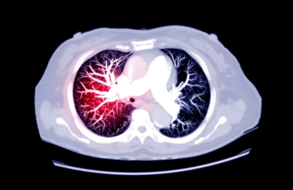
Pulmonary embolism occurs when a blood vessel is blocked in your lungs. It needs to be treated quickly because it is life-threatening.
When blood is present outside the blood vessel, probably following trauma to the blood vessel, it is the innate response of the body to stop the bleeding and further loss of blood. This is called hemostasis – a process that involves multiple interlinked four steps that occur in a rapid sequence.
Firstly, the blood vessel goes into spasms (Vasoscular spasms) causing narrowing of the blood vessel thus reducing blood flow and blood loss. In the second step, platelets stick together to form a temporary seal to cover the break in the vessel wall. This is called the platelet plug formation.
Thirdly once the platelet plug has been formed by the platelets, there is an activation of a sequence of events known as the ‘coagulation cascade’ which leads to the fourth stage, the formation of fibrin around the platelet plug to hold it in place, this makes the platelet plug to become harder. The resultant plug is called a ‘thrombus’ or colloquially called blood ‘Clot’.
This is often a good step for wound healing although, it can cause severe health problems if the thrombus becomes detached from the wall of the blood vessel and travels through the circulatory system with catastrophic complications of obstructing blood flow to the tissues supplied by the occluded blood vessels leading to restriction of blood flow and even tissue death.
In this way, a physiologic process becomes a pathologic process leading to morbidity and or mortality. If the thrombus reaches the lungs it could lead to pulmonary embolism or Stroke and heart attack if it gets to the brain and heart respectively.
However, without this process, the healing of a wound will not be possible. At times, this process is triggered inadvertently while the blood is within the lumen of the blood vessel and without any bleeding. Please also note the difference between a thrombus and an embolus.
A thrombus is a blood clot that forms in a vein while an embolus is anything that moves through the blood vessels until it reaches a vessel that is too small to let it pass.
What is Pulmonary Embolism?
Pulmonary embolism (PE) results when a thrombus, usually originating in the deep venous system of the lower extremities or rarely from the pelvic, renal, upper extremity veins, or the right heart chambers dislodge and travels through the venous system to lodge in the pulmonary arteries, where they partially or completely occlude one or more vessels.
The consequences depend on:
- The size and number of emboli.
- The underlying condition of the lungs.
- How well the right ventricle (RV) of the heart is functioning.
- The ability of the body’s intrinsic thrombolytic system to dissolve the clots.
Small emboli may have no acute physiologic effects and may begin to lyse immediately and resolve within hours or days while large thrombi can lodge at the bifurcation of the main pulmonary artery (Saddle pulmonary embolism) or the lobar branches and cause hemodynamic compromise.
Pulmonary embolism can also arise from non –thrombotic sources (e.g., embolism of air, amniotic fluid, fat, infected material, foreign body, tumour).
When a large embolus acutely occludes major pulmonary arteries or when many smaller emboli combine to occlude > 50% of the more distal lung blood vessels, RV pressure increases resulting in acute right ventricular failure, shock, or sudden death.
Death occurs due to right ventricular failure.

Risk Factors of Pulmonary Embolism (PE)
- Conditions that impair venous return, include bed rest and confinement without walking.
- Conditions that cause endothelial injury or dysfunction.
- Underlying inherited or acquired condition that increases the risk of excessive blood clot formation such as cancer or primary clotting disorders.
- Covid -19 appears to increase the risk for deep venous thrombosis and pulmonary embolism. Although part of the risk may be due to reduced mobility associated with illness.
Who is at Risk of Developing a Blood Clot?
People at risk for developing a blood clot are those who:
- Have been inactive or immobile for long periods due to bed rest, surgery or while travelling via motor vehicle, train or plane.
- With a personal or family histories of a blood clotting disorder, such as deep vein thrombosis (DVT) or pulmonary embolism (PE).
- Have a history of cancer or are receiving chemotherapy.
- Have a history of heart failure or stroke.
- Are overweight or obese
- Have recently had trauma or injury to a vein, possibly after a recent surgery, fracture or due to varicose veins.
- Are pregnant or have given birth in the previous 6 weeks.
- Are taking birth control pills (oral contraceptives) or hormone replacement therapy.
- Placement of central venous catheters through the arm or leg
Pathophysiology
Larger emboli can cause:
- A reflex increase in breathing (tachypnea),
- Low oxygen levels in the blood due to ventilation/perfusion (V/Q) mismatch and low mixed venous oxygen content as a result of low cardiac output.
- Partial or complete collapse of the entire lung area due to reduced alveolar carbon dioxide levels.
- Abnormalities in the lung surfactant (chemicals that keep the lungs from collapsing) and an increase in pulmonary vascular resistance caused by mechanical obstruction and vasoconstriction result in increased heart rate and low blood pressure.
Endogenous lysis reduces most emboli, even those of moderate size, and physiologic alterations decrease over hours or days. Some emboli resist lysis and may organize and persist and sometimes cause chronic pulmonary hypertension
Classification of Pulmonary Embolism
Pulmonary emboli may be classified according to the physiologic effects as
- High risk (catastrophic or super-massive): Here, the right ventricular function is impaired with severely low blood pressure / low oxygen level in the body.
- High risk (massive): The right ventricular function is also impaired causing low blood pressure.
- Intermediate risk (submassive): Also here, the right ventricular function is impaired with abnormal troponin and/or brain (B-type) natriuretic peptide (BNP) but no low pressure.
- Low risk: Absence of right ventricular impairment and absence of low blood pressure.
Symptoms of Pulmonary Embolism
Symptoms of pulmonary embolism vary, depending on the severity of the clot. Although most people with a pulmonary embolism experience symptoms, some will not. The first signs are usually shortness of breath and chest pains that get worse on exertion. There may cough with bloody sputum.
Symptoms may include:
- Sudden shortness of breath — whether you’ve been active or at rest.
- Unexplained sharp pain in your chest, arm, shoulder, neck or jaw. The pain may also be similar to symptoms of a heart attack.
- Cough with or without bloody sputum (mucus).
- Pale, clammy or bluish-coloured skin.
- Rapid heartbeat (pulse).
- Altered mental status.- especially in elderly patients.
- Excessive sweating.
- In some cases, feel anxious, light-headed, faint or passing out.
- Wheezing
- Low-grade fever
- Pain, swelling, and/or erythema of a leg or an arm)- signs of deep vein thrombosis
The most common signs of pulmonary embolism are:
- Increased heart rate (Tachycardia)
- Fast breathing (Tachypnea)

Investigations / Diagnosis of Pulmonary Embolism
The initial evaluation should include
-
Pulse oximetry
Provides a quick way to assess oxygenation; low oxygen concentration is one sign of PE, and it requires further evaluation.
-
Chest x-ray
Chest X-ray but may show
- Lung collapse (Atelectasis).
- Focal infiltrates.
- An elevated hemi diaphragm.
- Pleural effusion.
The classic findings of focal loss of vascular markings (Westermark sign), a peripheral, wedge-shaped density arising from the pleura (Hampton hump
A chest x-ray can also help exclude pneumonia(Pulmonary infarction due to pulmonary embolism may be mistaken for pneumonia)
-
ECG
Most often shows tachycardia and various ST wave abnormalities, which are not specific for pulmonary embolism
-
D-dimer testing
Elevated levels occur in the presence of a recent thrombus. A negative D-dimer test is highly indicative of the absence of pulmonary embolism.
However, elevated D-dimer levels are not specific for venous thrombus because many patients without deep venous thrombosis (DVT) or PE also have elevated levels (particularly in the inpatient setting).
Therefore, further testing is required when the D-dimer level is elevated. Very high D-dimer levels appear to predict a poor outcome.
-
The Pulmonary Embolism Rule-Out Criteria (PERC) rule
The PERC rule specifies 8 criteria. The presence of these criteria in a clinically low-risk patient specifies that testing for PE is not indicated. The criteria are:
- Age < 50 years
- HR < 100
- Oxygen saturation ≥ 95%
- No prior deep venous thrombosis or pulmonary embolism
- No unilateral leg swelling
- No estrogen use
- No hemoptysis
- No surgery or trauma requiring hospitalization within the past 4 weeks
Use of the PERC rule has been recommended as a way to decrease rates of testing for PE with conventional testing using D-dimer, but with similar rates of sensitivity and negative predictive values.
-
CT angiography
Is the preferred imaging technique for diagnosing acute pulmonary embolism. It is rapid, accurate, highly sensitive and specific.
It can also give more information about other lung pathology (e.g., demonstration of pneumonia rather than PE as a cause of hypoxia or chest pain) as well as the severity of PE (for example by the size of the right ventricle or the reflux into the hepatic veins
-
Ventilation/perfusion (V/Q) scans
This detects areas of the lung that are ventilated but not perfused. V/Q scanning takes longer than CT angiography and is less specific. It is particularly useful when contrasts required for CT- angiography cannot be used due to poor renal functions.
In some hospitals, V/Q scanning can be done with a portable machine that provides 3 views of ventilation and perfusion, which is useful when a patient is too ill to move.
-
Duplex ultrasonography
Is a safe, noninvasive, portable technique for detecting leg or arm thrombi. A clot can be detected by showing poor compressibility of the vein or by showing reduced flow by Doppler ultrasonography. Confirming DVT in the calf or iliac veins can be more difficult but can generally be accomplished. In suspected acute pulmonary embolism, the absence of venous thrombosis on ultrasonography does not rule out PE.

-
Echocardiography
This may show a clot in the right atrium or ventricle, but echocardiography is most commonly used for risk stratification in acute PE. The presence of right ventricular dilation and reduced ventricular range of movements may suggest the need for more aggressive therapy.
-
Cardiac marker testing
This method is evolving as a useful means of stratifying mortality risk in patients with acute pulmonary embolism. Cardiac marker testing can be used as an adjunct to another testing if PE is suspected or proven.
Elevated troponin levels signify right ventricular (or sometimes left ventricular) decreased blood supply.
Elevated brain natriuretic peptide (BNP) and pro-BNP levels may signify RV dysfunction; however, these tests are not specific for RV dysfunction or PE.
-
Thrombotic disorder (thrombophilia)
Testing should be considered for patients with PE and no known risk factors, especially if they are younger, have recurrent PE, or have a positive family history.
-
Pulmonary arteriography
Is now rarely needed to diagnose acute PE because noninvasive CT angiography has similar sensitivity and specificity. However, in patients in whom catheter-based thrombolytic therapy is being used, pulmonary angiography is useful for the assessment of catheter placement and may be used as a rapid means of determining the success of the procedure when the catheter is removed. Pulmonary arteriography is also still used together with right-heart catheterization in assessing whether patients with chronic thromboembolic pulmonary hypertension are candidates for pulmonary endarterectomy.
Treatment for Pulmonary Embolism
In many well-established centres, the management and treatment of PE are undertaken by a group of multi-disciplinary teams- comprising clinicians in pulmonary/critical care medicine, interventional cardiology, cardiothoracic surgery and haematology.
They rapidly evaluate and risk-stratify patients with pulmonary embolism and make the complex treatment decisions needed.
-
Supportive Therapy
- In patients with reduced oxygen supply, oxygen should be given.
- In patients with low blood pressure due to massive PE, 0.9% saline can be cautiously given IV. Overloading the right ventricle can result in deterioration.
- Drugs that constrict blood vessels may also be given if IV fluids fail to sufficiently increase blood pressure. Norepinephrine is the most commonly used first-line
- Hospitalization for at least 24 to 48 hours is done for most patients with PE. Patients with abnormal vital signs or massive or sub-massive PE require longer periods of hospitalization.
- ICU admission is always required for massive PE. ICU admission should also be considered if patients have:
- Extensive clot burden
- Right ventricular compromise
- Significant low oxygen blood levels
- Low or borderline blood pressure
- Clinical deterioration
-
Anticoagulation
This is the mainstay of therapy for PE, and rapid reduction of clot burden using thrombolytic therapy or embolectomy is indicated for patients with:
- Low blood pressure that does not resolve after fluid resuscitation,
- Selected patients with impaired right ventricular function
Initial anticoagulation followed by maintenance anticoagulation is indicated for patients with acute pulmonary embolism to prevent clot extension and further embolization as well as new clot formation.
Anticoagulant therapy for acute PE should be started whenever PE is strongly suspected, as long as the risk of bleeding is deemed low. Initial anticoagulation choices for acute PE include
-
Intravenous unfractionated heparin
- Has a short half and is reversible with Protamine.
- Requires ongoing hospitalization to administer.
- Regardless many clinicians prefer this intravenous unfractionated heparin regimen.
-
Subcutaneous low molecular weight heparin
Has several advantages over unfractionated heparin including:
-
- Superior bioavailability.
- Weight-based dosing results in a more predictable anticoagulation effect than does weight-based dosing of unfractionated heparin.
- Ease of administration (can be given subcutaneously once or twice a day)
- Decreased incidence of bleeding.
- Potentially better outcomes.
- The potential for patients to self-inject (thereby allowing earlier discharge from the hospital).
- Low molecular weight heparins are generally contraindicated in patients with severe renal insufficiency
- Low molecular weight heparins are partially reversible with protamine.
-
Subcutaneous fondaparinux
This is a factor Xa antagonist given subcutaneously. It can be used in acute deep vein thrombosis and acute PE instead of heparin or low molecular weight heparin.
It has also been shown to prevent recurrences in patients with superficial venous thrombosis. Outcomes appear to be similar to those of unfractionated heparin.
-
Inferior vena cava filter placement (in selected patients)
Placement of a removable percutaneous inferior vena cava filter (IVCF) should be considered for patients with:
- Contraindications to anticoagulation.
- for those with recurrent PE despite anticoagulation.
-
Rapid clot burden reduction (in selected patients)
-
Thrombolytic therapy
Thrombolytic medications (“clot busters”), including tissue plasminogen activator (TPA), are used to dissolve the clot. Thrombolytic are always given in a hospital where the patient can be closely monitored. These medications are used in special situations, such as if the patient’s blood pressure is low or if the patient’s condition is unstable due to pulmonary embolism.
-
Catheter-directed therapy
Catheter-directed PE therapy (thrombolytics, embolectomy) uses catheter placement in the pulmonary arteries for disruption and/or lysis of the clot.
Maintenance of Anticoagulation for Pulmonary Embolism
This is indicated to reduce the risk of clot extension or embolization and reduce the risk of new clot formation. Drug choices for maintenance anticoagulation include
- Oral vitamin K antagonist (warfarin in the US)
- Oral factor Xa inhibitors (Rivaroxaban)
- Oral direct thrombin inhibitor (dabigatran
Warfarin is an effective long-term oral anticoagulant option that has been used for decades, but it is very inconvenient for several reasons. In most patients, Warfarin is started on the same day as heparin (or fondaparinux therapy used for initial anticoagulation. heparin (or fondaparinux therapy should be overlapped with Warfarin therapy for a minimum of 5 days and until the INR has been within the therapeutic range (2.0 to 3.0) for at least 24 hours.
The major disadvantages of warfarin are the need for periodic INR monitoring, frequent dose adjustments, and drug interactions.
The oral factor Xa inhibitor anticoagulants, Rivaroxaban can be used for both initial and maintenance anticoagulation therapy These drugs are more convenient than warfarin due to their fixed dosing and lack of need for laboratory monitoring, as well as having fewer drug
Duration of Maintenance
Anticoagulation for PE is dependent on a variety of factors:
- Risk factors for PE and bleeding risk can range from 3 months to lifelong therapy.
- Transient risk factors (e.g., immobilization, recent surgery, trauma) require only 3 months of treatment.
- Patients with unprovoked PE, those with more durable risk factors for PE (e.g., cancer, thrombophilic disorder), and those with recurrent PE might benefit from lifelong anticoagulation provided the bleeding risk is low or moderate

How Do I Prevent Pulmonary Embolism?
- Exercise regularly. If you can’t walk around due to bed rest, recovery from surgery or extended travel, move your arms, legs and feet for a few minutes each hour. If you know you will need to sit or stand for long periods, wear compression stockings to encourage blood flow.
- Drink plenty of fluids, like water and juice, but avoid excess alcohol and caffeine.
- If you need to be stationary for long periods, move around for a few minutes each hour: move your feet and legs, bend your knees, and stand on tiptoe.
- Do not smoke.
- Avoid crossing your legs.
- Do not wear tight-fitting clothing.
- Lose weight if you are overweight.
- Elevate your feet for 30 minutes twice a day
- Talk to your doctor about reducing your risk factors, especially if you or any of your family members have experienced a blood clot.
- A high-risk patient may use anticoagulant drugs such as heparin or warfarin.
- Compression of the legs is possible, using anti-embolism compression stockings or pneumatic compression. An inflatable sleeve, glove, or boot holds the affected area and increases the pressure when required.
Conclusion
Pulmonary embolism is a serious but very treatable condition. A quick treatment greatly reduces the chance of death. An estimated 10% of patients with pulmonary embolism die within the first few hours after presentation.
Most patients who die as a result of acute PE are never diagnosed before death. PE is not suspected in most of these patients. The best prospects for reducing mortality involve
- Improving the frequency of diagnosis (e.g., by including PE in the differential diagnosis when patients present with nonspecific but compatible symptoms or signs.
- Improving the rapidity of diagnosis.
- Improving risk-stratification.
- Improving the rapidity of initiation of anticoagulation therapy.
- Providing appropriate prophylaxis in at-risk patients.