Molar Pregnancy is an uncommon pregnancy complication characterized by abnormal growth of trophoblasts, which are the cells that typically develop into the placenta. How does normal fertilisation happen? Fertilization is the union of a sperm nucleus of paternal origin, with an egg nucleus of maternal origin to form the primary nucleus of an embryo.
In all organisms, the essence of fertilization is the fusion of the hereditary material of two different sex cells or gametes each of which carries half the number of chromosomes typical of the species. In humans, chromosomes come in pairs. Normally, each cell in the human body has 23 pairs of chromosomes (a totally of 46 chromosomes in all). Half comes from the mother while the other half comes from the father.
Two of the chromosomes (X and Y chromosomes)are called the sex chromosomes and they determine the sex of the child as male or female. Females have 2 X chromosomes and males have 1 X and 1 Y chromosome while 22 chromosomes are called autosomal chromosomes because they are the same in both males and females.
The mother contributes an X chromosome, while the father contributes either an X or a Y. The chromosome from the father determines the sex.
In a molar pregnancy, cytogenic studies have shown that 90% of them have a 46XX (diploid) pattern, all derived from the sperm that replicates after fertilization. This phenomenon is called androgenesis. They are presumed to be derived from fertilization by a single sperm of an egg that has lost its chromosome (empty egg) and the remaining 10% from the fertilization of such an empty egg by 2 sperm. In both circumstances, the embryo dies early and thus, complete moles show no fetal parts. Here we have triploid pattern 69xxy.
What is a Molar Pregnancy?
It is an abnormal pregnancy, one of the spectrums of proliferative diseases called gestational trophoblastic disease (GTD)of which are 3 types of GTD viz
- Hydatiform moles
They are benign placental tumours with malignant potential.
- Gestational trophoblastic neoplasia
- Malignant placental tumours.
For this article, we will be considering Hydatiform moles. It is a fertilization error, which arises when an ovum without maternal chromosomes (empty egg) is fertilized by one sperm which then duplicates its DNA, resulting in a situation in which all the chromosomes are paternally derived or arising from fertilization of an “empty ovum” by two sperm.
Most (> 80%) hydatidiform moles are benign( not cancerous). In patients with a prior partial or complete mole, the incidence of a second mole in subsequent pregnancies is 1 to 2%. Patients who have had a mole require ultrasonography early in subsequent pregnancies, and the placenta should be sent for pathology evaluation. Patients with consecutive molar pregnancies require germline genetic testing for mutations.
Types of Hydatiform moles
Two types are known viz:
-
Complete Mole
- Fertilization of an ovum whose nucleus is either absent or inactivated (empty egg) by a sperm which then duplicates.
- All placentavilla swelled.
- Fetus, cord, amniotic fluid, and membranes are absent.
- Paternal chromosomes only (46xx).
- Diploidy.
-
Partial mole
- Fertilization of an ovum whose nucleus is either absent or inactivated (empty egg) by two sperms.
- Some placenta villa is swollen.
- Fetus, cord, amniotic fluid, and membranes are present.
- Paternal and Maternal chromosomes (69xxy).
- Triploidy.
Predisposing Factors for Molar Pregnancy

- Extremes of maternal age
Both advanced and very young maternal age has consistently correlated with higher rates of complete mole. These observations suggest that ova of very young or older women are predisposed to abnormal fertilization events that lead to complete hydatiform moles.
Importantly, the association between maternal age and complete molar pregnancies is not seen in women with partial molar pregnancies
- Prior molar pregnancy
Prior complete molar pregnancy increases the risk of developing a subsequent complete molar pregnancy. The risk of a repeat molar pregnancy after one mole is approximately 1%, while after two moles; the risk of a third mole is 15% to 20%.
- History of prior spontaneous abortion. It also appears to increase the risk of molar pregnancy (both complete and partial).
- Dietary deficiency. Deficiency in β-carotene and animal fat has been linked to an increase in complete moles.
- history of oral contraceptive use. There appears to be a possible increased risk of molar pregnancy (partial and complete) with a history of oral contraceptive use, while ovulation induction regimens may be associated with an increase in twin pregnancies consisting of a normal fetus (es) and a complete mole.
Furthermore, partial molar pregnancies are more common in women with a history of irregular menses, miscarriage, and oral contraceptive use for > 4 years, but are not associated with ethnicity, ovulation induction, or dietary factors.
Symptoms of Molar Pregnancy
- Initial manifestations of a hydatiform mole suggest early pregnancy with a positive pregnancy test. But because molar pregnancies grow much faster than a fetus, the abdomen becomes larger much faster than it does in a normal pregnancy.
- Vaginal bleeding.
- Severe nausea and vomiting (excessive vomiting in pregnancy).
- Absent fetal movement and fetal heart sounds.
- As parts of the mole deteriorate, small amounts of tissue, which resemble a bunch of grapes, may pass through the vagina. This symptom indicates the need for prompt evaluation by a doctor and strongly suggests the diagnosis.
Molar Pregnancy Diagnosis/Investigations
-
Symptoms of molar pregnancy
Often, molar pregnancy (hydatiform mole) can be diagnosed shortly after it forms. It is suspected based on symptoms, such as a uterus that is much larger than expected and a vaginal discharge of grapelike tissues
-
Blood tests
- A pregnancy test is done and the results are usually positive.
- Blood tests to measure the level of human chorionic gonadotropin (HCG—a hormone normally produced early in pregnancy) are done. In molar pregnancy or another type of gestational trophoblastic disease is present, the level is usually very high because these tumours produce a large amount of this hormone.
The level of human chorionic gonadotropin in the blood is measured to determine whether the molar pregnancy was completely removed. When removal is complete, the level returns to normal, usually within 10 weeks, and remains normal, and no further treatment is needed.
While HCG is being measured, women should use effective contraception because pregnancy makes interpreting hCG measurement difficult. If the level does not return to normal, the disease is considered persistent
- Thyroid function Test (TFT)
If the level of HCG is very high, blood tests to check thyroid function are done and determine whether hyperthyroidism (overactive thyroid gland) is present. This can occur in women with hydatiform moles or other Gestational trophoblastic diseases. Symptoms of which include:
-
- Abnormally fast heart rate (tachycardia).
- Warm skin.
- Sweating
- Heat intolerance.
- Mild tremors.
-
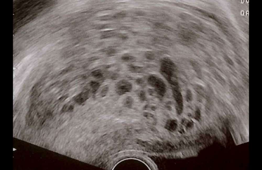
Imaging
- Ultrasonography

Ultrasonography is done to be sure that the growth is a hydatiform mole and not a fetus or amniotic sac (which contains the fetus and fluid around it).
No fetal movement and no fetal heartbeat are detected
- CT- Chest, abdomen and pelvis or Chest x-ray
If Hydatiform mole or other gestational trophoblastic disease is diagnosed, tests are done to find out if there is a spread from where it started to other parts of the body. Tests include computed tomography (CT) of the chest, abdomen, and pelvic area. MRI may also be done.
-
Biopsy
Samples of tissue removed during a D &C or obtained when tissue is passed is then examined under a microscope (biopsy) to confirm the diagnosis.
Treatment of Molar Pregnancy
-
Removal of the mole
A molar pregnancy (hydatidiform mole) or any type of gestational trophoblastic neoplasia is removed completely usually by dilatation and curettage (D and C) or with suction aspiration.
-
Chemotherapy
Chemotherapy is needed if the mole persists or has spread. If the mole is considered low risk, chemotherapy may consist of only one drug (methotrexate or dactinomycin). If this treatment is ineffective, a combination of chemotherapy drugs (such as etoposide, methotrexate, dactinomycin, cyclophosphamide, and vincristine) may be used.
-
Surgery
Removal of the uterus is rarely necessary but may be done for women who do not plan to have children and if chemotherapy is not effective.
-
Combination treatment (chemotherapy, radiotherapy and surgery)
If the mole has spread widely and is considered high risk, referral to a specialist is advised, here treatment usually involves several chemotherapy drugs, radiation therapy, as well as the removal of the uterus (hysterectomy)
After a hysterectomy, if done, chemotherapy is given and hCG levels are monitored to make sure the disease has been successfully treated.
Post Treatment of Molar Pregnancy
When the gestational trophoblastic disease is diagnosed, doctors talk to women about their desire to be able to have children usually, any type of gestational trophoblastic disease can be successfully diagnosed and treated, without endangering reproductive function.
Women who have had a molar pregnancy removed are advised not to become pregnant for I year, during this period, effective contraceptive methods can be used. Pregnancy is delayed so that doctors can make sure that treatment was successful.
If women who have had a molar pregnancy become pregnant, early ultrasound should be done to determine whether the pregnancy is normal. After the baby is delivered, the placenta is usually sent to a histopathology laboratory abnormality check.
Follow-up requires serial serum HCG measurements every 1 to 2 weeks until three consecutive tests show normal HCG levels, after which HCG levels should be determined at 3-month intervals for 6 months after the spontaneous return to normal. Contraception is recommended during this follow-up period for 6 months after the first normal HCG result.
Oral contraceptives are preferred because they have the advantage of suppressing endogenous luteinizing hormone (LH), which may interfere with the measurement of HCG at low levels, and does not increase the risk of post molar GTN.
Conclusion
Most women who have had a molar pregnancy can have children afterwards and do not have a higher risk of a miscarriage, complications during pregnancy, or children with birth defects.
About 1 to 2% of women who have had a molar pregnancy will have another one. So if a woman has had a molar pregnancy, an ultrasound scan is done early in a subsequent pregnancy. If she has consecutive molar pregnancies, genetic testing is done.
Medical induction of labour and hysterectomy is not recommended for molar evacuation. These methods increase maternal morbidities, such as blood loss, incomplete evacuation requiring curettage, and the requirement for cesarean delivery in subsequent pregnancies.
They also increase trophoblastic dissemination and the development of postmolar GTN requiring chemotherapy.