To understand gout, it is important to know about Purines and their metabolism. Purines are nitrogen-containing compounds that come from the food we eat and from the breakdown of nucleic acid in the body (the major site of purine nucleotide synthesis is in the liver).
In as much as purine is one of two chemical compounds that cells use to make the building blocks of DNA and RNA, excessively high levels can lead to health problems, including gout and hyperuricemia (excessive levels of uric acid in the blood).
Purines also act as metabolic signals, provide energy, control cell growth, are part of essential coenzymes, contribute to sugar transport and donate phosphate groups in phosphorylation reactions.
High-purine foods are also high-protein food but proteins are not purine because they have a completely different chemical structure.
Dietary purines are present in almost all foods. However, certain foods have high levels of purines, and one should eat in moderation if one has gout or hyperuricemia.
Foods that are very high in purine include:
- Anchovies.
- Beef.
- Goose.
- Herring.
- Mackerel.
- Mussels.
- Organ meats (such as liver and kidney).
- Yeast.
Among foods with moderate levels of purines are other fish, meat, poultry and shellfish, asparagus, beans, lentils, dried peas and spinach.
When the body breaks down purine, a chemical called uric acid is produced. Most uric acid dissolves in blood and travels to the kidneys where it is passed out in urine.
If the body produces too much uric acid or does not remove enough of it, a high level of uric acid accumulates in the blood (hyperuricemia).
Humans and higher-order primates tend to retain high levels of serum uric acid due to the lack of a key enzyme, uricase, which converts uric acid to water-soluble allantoin as in lower animals.
When there is too much uric acid in the body, uric acid crystals (monosodium urate, MSU) build up in joints, fluids, and tissues within the body thus leading to gout.
Please, note hyperuricemia does not always cause gout (hyperuricemia without gout symptoms does not need to be treated!).
What is gout?
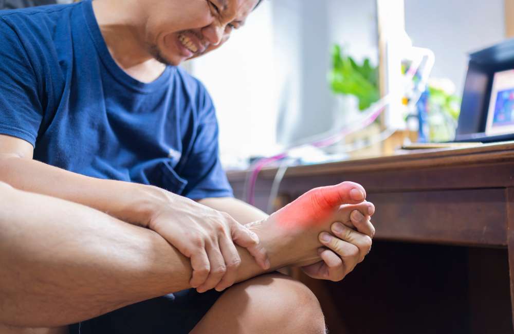
A gout is a common form of inflammatory arthritis that is very painful. It usually affects one joint at a time (often the big toe joint). Repeated bouts of gout can lead to gouty arthritis, a worsening form of arthritis. Symptoms get worse occasionally (known as ‘flares’, and when there are no symptoms, it is called ‘remission’.)
There is no cure for gout, but can effectively be treated and managed with medication and self-management strategies. Please note that pseudo gout is caused by calcium pyrophosphate crystals and is more accurately termed calcium pyrophosphate disease while gout is caused by monosodium urate monohydrate crystals.
The symptoms of pseudogout are very similar to those of gout, although the flare-ups are usually less severe. Monosodium urate (MSU) crystals which precipitate as needle-shaped crystals, are deposited extracellularly in avascular tissues (e.g., cartilage) or in relatively avascular tissues (e.g., tendons, tendon sheaths, ligaments, walls of bursae etc.) and skin around cooler distal joints and tissues (e.g., ears, finger pads).
In severe, long-standing hyperuricemia, MSU crystals may be deposited in larger central joints and the parenchyma of organs such as the kidney. At the acid pH of urine, urate precipitates readily as small plate-like or diamond-shaped uric acid crystals that may aggregate to form gravel or stones which may obstruct urine outflow.
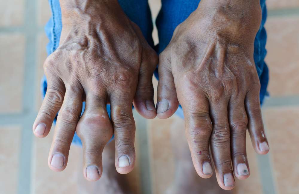
Tophi are MSU crystal aggregates that most often develop in joint and cutaneous tissue. They are usually encased in a fibrous granulomatous matrix, which prevents them from causing acute inflammation.
Causes of gout
Urate levels can be elevated because of viz:
-
Decreased renal (most common) or gastrointestinal excretion.
This is by far the most common cause of high levels of uric acid accumulation in the blood. It may be:
-
- Hereditary e.g. When there are variations in uric acid transport system. Also decreased renal excretion occurs in patients receiving diuretics and in patients suffering from disease conditions that decrease the glomerular filtration rate
- Ethanol
Ethanol increases purine breakdown in the liver and increases the formation of lactic acid which blocks urate secretion by the renal tubules and also stimulates liver urate production.
-
- Lead poisoning/High dose of cyclosporine
Lead poisoning and high doses of cyclosporine given to renal transplant patients alter renal tubular function thus causing urate to be retained.
-
Increased production (rare).
Increased production of urate may be caused by:
-
- Increased nucleoprotein turnover in hematologic conditions (e.g., lymphoma, leukaemia, hemolytic anaemia).
- In conditions with increased rates of cellular proliferation and cell death (e.g., psoriasis, cytotoxic cancer therapy, radiation therapy).
- Primary hereditary abnormality and in obesity, because urate production correlates with body surface area.
- Enzyme abnormalities can be possible causes of increased urate production viz deficiency of hypoxanthine-guanine phosphoribosyl transferase or over activity of phosphoribosyl pyrophosphate synthetase.
-
Increased purine intake (usually in combination with decreased excretion).
Increased intake of purine-rich foods (e.g., liver, kidney, anchovies, asparagus, consommé, herring, meat gravies and broths, mushrooms, mussels, sardines, sweetbreads) can contribute to hyperuricemia. Beer, including nonalcoholic beer, is particularly rich in guanosine, a purine nucleoside.
How does gout progress?
There are various stages of gout progression viz:
- Asymptomatic hyperuricemia.
A person can have elevated uric acid levels without any outward symptoms. These individuals do not need treatment at this stage. High uric acid levels in the blood can cause silent tissue damage. In situations like this, factors possibly contributing to its buildup are addressed.
- Acute gout.
This stage occurs when urate crystals in a joint suddenly cause acute inflammation and intense pain. This sudden attack is called a “flare” and may last between 3-13 days. Stressful life events and excessive alcohol consumption could be contributors to flare-ups.
- Interval or intercritical gout.
This stage is the period in between attacks of acute gout. As a person’s gout progresses, these intervals become shorter. Between these periods, urate crystals may continue to build up in tissue.
- Chronic tophaceous gout.
Chronic tophaceous gout is the most debilitating type of gout and may result in permanent damage to the joints and the kidneys. At this stage, people can have chronic arthritis and develop tophi in cooler areas of the body, such as the joints of the fingers.
Chronic tophaceous gout typically occurs after many years of acute gout attacks. However, it is unlikely that individuals who receive proper treatment progress to this stage.
What increases the chances of gout (risk factors)?
The following increase the likelihood of hyperuricemia and gout viz:
- Age.
Gout is more common in older adults and rarely affects children.
- Sex.
Being male is a risk factor. In people under the age of 65 years, gout is 4 times as prevalent among males than females.
- Genetics.
A family history of gout can increase the likelihood of a person developing the condition.
- Lifestyle choices.
-
- Alcohol consumption interferes with the removal of uric acid from the body.
- Eating a high-purine diet also increases the amount of uric acid in the body. Both of these can lead to gout.
- Eating or drinking food and drinks high in fructose (a type of sugar).
- Lead exposure.
Studies have suggested a link between chronic lead exposure and an increased risk of gout.
- Medications.
Certain medications can increase the levels of uric acid in the body. These include some diuretics and drugs containing salicylate.
- Weight
Being overweight or obese and having high levels of visceral body fat have associations with an increased risk of gout. However, being overweight or obese cannot directly cause the condition.
- Having certain health conditions, including:
-
- Congestive heart failure.
- Hypertension (high blood pressure).
- Insulin resistance.
- Metabolic syndrome.
- Diabetes.
- Poor kidney function.
- Renal insufficiency and other kidney conditions can reduce the body’s ability to remove waste, leading to elevated uric acid levels.
Signs/symptoms
- Joint Pain
The main symptom of gout is intense joint pain that subsides to discomfort, inflammation, and redness.
Acute gouty arthritis usually begins with sudden onset of pain (often at night). The metatarsophalangeal joint of a great toe is most often involved but the ankle, knee, wrist, and elbow are also common sites (rarely, the hip, shoulder, sacroiliac, sternoclavicular, or cervical spine joints are involved).
The pain becomes progressively more severe, usually over a few hours, and is often excruciating.
- Swelling, warmth, redness, and exquisite tenderness may suggest infection. The overlying skin may become tense, warm, shiny, and red or purplish.
- Fever and chills.
- Tachycardia (heart rate of over 100beats per minute).
- Malaise (General feeling of being unwell).
Investigations for gout
-
Synovial fluid analysis.
If acute gouty arthritis is suspected, arthrocentesis (a procedure where the doctor uses a needle to take fluid out of a joint) and synovial fluid analysis should be done at the initial presentation.
Every attempt should be made to document the presence of MSU crystals in synovial fluid from an affected joint.
-
Serum urate level.
An elevated serum urate level supports the diagnosis of gout but is neither specific nor sensitive; at least 30% of patients have a normal serum urate level during an acute flare.
The serum urate level can be low after the initiation of therapy, but flares can continue to occur as long as tissue deposits remain. The dissolution of the urate deposits may take many months after initiating therapy.
-
Imaging.
-
- X-rays
X-rays of the affected joint may be taken to look for bony erosions or tophi. X-rays cannot detect gout, but healthcare professionals may use them to rule out other causes and are unnecessary if the diagnosis of acute gout has been established by synovial fluid analysis and rarely show erosions at the time of first flares.
-
- Ultrasound scan
Ultrasonography is more sensitive for the diagnosis of gout. Urate deposition over the articular cartilage (double-contour sign) and clinically inapparent tophi are characteristic changes. These findings may be evident even before the first gout flare.
-
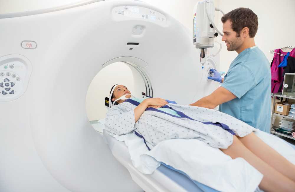
- CT-scan
Dual-energy CT scans (DECTs) can also reveal uric acid deposits and can be useful if the diagnosis is unclear based on standard clinical evaluation and testing, particularly if synovial fluid aspiration and analysis cannot be done.
Diagnosis
Gout can often be challenging to diagnose, as its symptoms are similar to those of other conditions. While elevated uric acid levels in the blood occur in most people who develop gout, it may not be present during a flare-up. As a result, a person does not need to have elevated uric acid levels in the blood for a diagnosis.
High levels of uric acid in an individual’s blood or urate crystals in their joint fluid are the main diagnostic for gout. So, to diagnose gout, one must have viz
- A high index of suspicion.
The diagnosis of gout should be suspected in patients with acute arthritis in one or multiple joints, particularly older adults or those with other risk factors. Affectation of the joint between the foot and the big toe and recurrent instep inflammation is particularly suggestive. Previous flares that began explosively and resolved spontaneously within 7 to 10 days are also characteristic.
- Results of the investigations stated above.
Can I protect myself from having gout?
Yes, one can prevent getting gout by doing the following:
- Maintaining a high fluid intake of around 2–4 liters a day.
- Avoiding alcohol.
- Maintaining a moderate weight.
Thus, the above prevention tips can be useful and can protect one against flares or prevent gout from occurring in the first instance.
Gout Treatment
-
Treatment of acute flare.
Termination of an acute flare with:
-
- Nonsteroidal anti-inflammatory drugs (NSAIDs).
They are effective in treating acute flares and are generally well tolerated. However, they can have adverse effects, including gastrointestinal upset or bleeding, hyperkalemia (low potassium), increases in creatinine, and fluid retention. Older and dehydrated patients are at particular risk, especially if there is a history of renal disease. Virtually any NSAID used in anti-inflammatory (high) doses is effective and is likely to exert an analgesic effect beginning within a few hours.
Treatment should be continued for several days after the pain and signs of inflammation have resolved to prevent relapse.
-
- Colchicine.
A traditional therapy that produces a dramatic response in some patients is begun soon after the onset of symptoms.
Renal insufficiency and drug interactions, especially with clarithromycin and some statins (drugs used for high cholesterol levels), may warrant a reduction of dosage or the use of other treatments. Gastrointestinal upset and diarrhea are common adverse effects.
If monotherapy is ineffective or doses (e.g., of NSAIDs) are limited by toxicity, Colchicine can be combined with NSAIDs or corticosteroids.
-
- Corticosteroids.
Corticosteroids are used to treat acute flares. Aspiration of affected joints, followed by the instillation of corticosteroid ester crystal suspension, is very effective, particularly for monarticular symptoms.
-
- Interleukin-1 (IL-1) antagonist
If corticosteroids, Colchicine, and NSAIDs are contraindicated or ineffective, an IL-1 antagonist, such as anakinra can be used. It hastens the resolution of a flare and shortens the hospital stay of a patient with multiple comorbidities that limit the use of the other drugs.
Anakinra is typically given as 100 mg subcutaneously once a day until symptoms resolve and it has the advantage of not affecting glucose levels or renal function or causing fluid retention and can be used in a patient with an active infection that is being appropriately treated.
-
Prevention of further deposition of monosodium urate (MSU) crystals, reduction in flare incidence, and resolution of existing tophi.
-
- Lowering the serum urate level by decreasing urate production with allopurinol or febuxostat (xanthine oxidase inhibitors (XOI).
Urate-lowering therapy is indicated for patients with the following viz:
-
- Tophaceous deposits.
- Evidence of joint damage due to gout on imaging studies.
- Frequent or disabling flares (e.g., more than 2 flares times a year) of gouty arthritis.
- Urolithiasis.
- Patients with infrequent flares but whose serum uric acid level of more than 9 mg/dL (more than 0.5 mmol/L) or for whom having any flares poses particular hardship.
- Multiple comorbidities (e.g., peptic ulcer disease, chronic kidney disease) that are relative contraindications to the drugs used to treat recurrent acute flares (NSAIDs or corticosteroids).
-
- Dissolving deposits with uricase replacement therapy.
- Increasing urate excretion with probenecid.
-
Prevention of recurrent acute flares.
This is done with daily Colchicine or an NSAID (if renal function allows).
-
Treatment of coexisting hypertension, hyperlipidemia, and obesity and avoidance of excess dietary purines.
The antihypertensive drug losartan and the triglyceride-lowering drug fenofibrate both have uricosuric effects and can be used to decrease uric acid in patients who have other reasons for taking these drugs.
-
The use of uricase
Pegloticase is a pegylated form of recombinant uricase. Uricase is an enzyme, absent in humans, that converts urate to allantoin, which is more soluble.
Pegloticase is expensive and is used primarily in patients with gout in whom other treatments have been unsuccessful in lowering the serum urate level and in patients who have a high burden of tophaceous deposits that would not likely be dissolved in a reasonable time period by other urate-lowering therapies.
It is given intravenously every 2 to 3 weeks for many months (typically at least 6 to 9 months) to totally deplete the excess urate deposits; it often lowers the serum urate level to less than 1 mg/dL (< 0.1 mmol/L). Pegloticase is contraindicated in patients with G6PD because it can cause hemolysis.
-
Other treatments
-
- Fluid intake of more than 3 L per day is desirable for all patients, especially those who chronically pass urate gravel or stones.
- Alkalization of urine with potassium citrate 20 to 40 mEq orally 2 times a day or acetazolamide 500 mg orally at bedtime) is also occasionally effective for patients with persistent uric acid urolithiasis despite hypouricemic therapy and adequate hydration.
However, excessive urine alkalization may cause the deposition of calcium phosphate and oxalate crystals.
- Extracorporeal shock wave lithotripsy may be needed to disintegrate renal stones.
- Large tophi in areas with healthy skin may be removed surgically; all others should slowly resolve under adequate hypouricemic therapy.
Complications of gout
Gouty arthritis can cause viz:
- Sever Joint pain with deformity and limited joint motion.
- Inflammation can be flaring in some joints while subsiding in others.
- Patients with gout may develop uric acid stones or calcium oxalate stones.
- Renal obstruction and infection, with the secondary tubulointerstitial disease.
- Untreated progressive renal dysfunction, most often related to coexisting hypertension or, less often, some other cause of nephropathy, further impairs the excretion of urate, accelerating crystal deposition in tissues.
Conclusion
Suspect gout if you have a sudden onset, unexplained single or joint pain involving a few joints, especially if the great toe or midfoot is affected, particularly if the pain keeps getting worse and you also have a high temperature (fever).
Please, notify your health practitioner to properly assess and confirm the diagnosis because other conditions that require urgent treatment, such as an infected joint, can sometimes cause similar symptoms.
Also note that although increased purine intake and increased production can contribute to increased uric acid in the blood, the most common cause of gout is decreased urate excretion due to kidney disorders or genetic variability in uric acid transport.
However, a strict low-purine diet lowers serum urate by only about 1 mg/dL (0.1 mmol/L) and thus is rarely sufficient therapy for patients with gout.