
Cataracts is a condition whereby the eye lens becomes cloudy. What’s the importance of the lens? The lens of the eye is a double convex, relatively acellular (not made up of cells or divided into cells), optically transparent intraocular structure that with the cornea serves to transmit light to the retina with minimal light scattering.
The main optical function of the lens is to transmit light, focusing it on the retina. The cornea contributes about 80% of total refraction, while the lens fine-tunes the focusing of light onto the retina.
The eye lens is made of transparent proteins called crystallins, hence the name crystalline lens. As one ages, the lenses in your eye become less flexible, less transparent and thicker. Age-related and other medical conditions cause these proteins and fibres within the lenses to break down and clump together, clouding the lenses.
In this defect, the crystalline lens becomes milky and cloudy. This condition is known as cataract and as the cataract continues to develop, the clouding becomes denser thus causing partial or complete loss of vision.
Removing the cataract through surgery, however, can restore this loss of vision.
What are Cataracts?
Cataracts is a congenital or degenerative opacity of the crystalline lens leading to gradual painless vision blurring.
Types of Cataracts
There are different types of cataracts classified based on where and how they develop in your eye viz:
-
Nuclear cataracts
This forms in the middle of the lens and causes the nucleus or the centre to become yellow or brown.
-
Cortical cataracts
This form around the edges of the nucleus and is wedge-shaped in appearance.
-
Posterior capsular cataracts
Affects the back of the lens and forms faster than the nuclear and cortical cataracts.
-
Congenital cataracts
These are present at birth or form during a baby’s first year. They are less common than age-related cataracts.
-
Secondary cataracts
Are caused by disease or medications. Diseases/medications that are linked with the development of cataracts include:
-
- Glaucoma.
- Diabetes.
- Uveitis
- The use of steroids (prednisone) and other medications can sometimes lead to cataracts.
-
Traumatic cataracts
Develop after an injury to the eye, but takes several years for it to develop.
-
Radiation cataracts
Results after radiation treatment for cancer.
What are the risk factors developing for cataracts?
Risk factors associated with cataracts include:
- Older age
Lens proteins denature and degrade over time, and this process is accelerated by diseases such (as diabetes, and hypertension), and environmental factors, including toxins, radiation, and ultraviolet light.

- Heavy alcohol use
Evidence is conflicting over the effect of alcohol. Some surveys have shown a link, but others, which followed people over a longer time, have not.
- Smoking
Has been shown to double the rate of nuclear sclerotic cataracts and triple the rate of posterior sub-capsular cataracts.
- Obesity
- Hypertension
- Previous eye trauma
Blunt trauma causes swelling, thickening, and whitening of the lens fibres. While the swelling normally resolves with time, the white colour may remain.
- Family history of cataracts
The genetic component is strong in the development of cataracts, most commonly through mechanisms that protect and maintain the lens.
- Chronic ultraviolet light exposure (sun exposure)
Ultraviolet light has been shown to cause cataracts, and some evidence indicates sunglasses worn at an early age can slow its development in later life.
- Undernutrition
- Diabetes
Blood sugar is the link between diabetes and cataracts. As the blood sugar rises, there is a high sugar level in the aqueous humour, the fluid between the eyeballs and the cornea which supplies nutrients and oxygen to the lens.
Uncontrolled blood sugar also causes enzymes in the lens to convert glucose to a substance called sorbitol. Too much sorbitol in the lens leads to cloudy vision.
- Exposure to radiation from X-rays and cancer treatments
Cataracts can arise as an effect of exposure to various types of radiation. X-rays, one form of ionizing radiation may damage the DNA of lens cells.
- The heat from infrared exposure
- Skin diseases
The skin and the lens have the same embryological origin and so can be affected by similar diseases. Those with atopic dermatitis and eczema occasionally develop shield ulcer cataracts.
- Post vitrectomy
Nearly every person who undergoes a vitrectomy without ever
having had cataract surgery will experience the progression of cataracts after the operation.
- Inadequate vitamin c intake
Low vitamin c intake and serum levels have been associated with greater cataract rates. However, the use of supplements of vitamin C has not demonstrated benefit
Symptoms of cataracts
Common symptoms of cataracts include:
- Blurry vision.
- Trouble seeing at night.
- Seecolourslors as faded.
- Increased sensitivity to glare.
- Halos surrounding lights.
- Double vision in the affected eye.
- A need for frequent changes in prescription glasses.
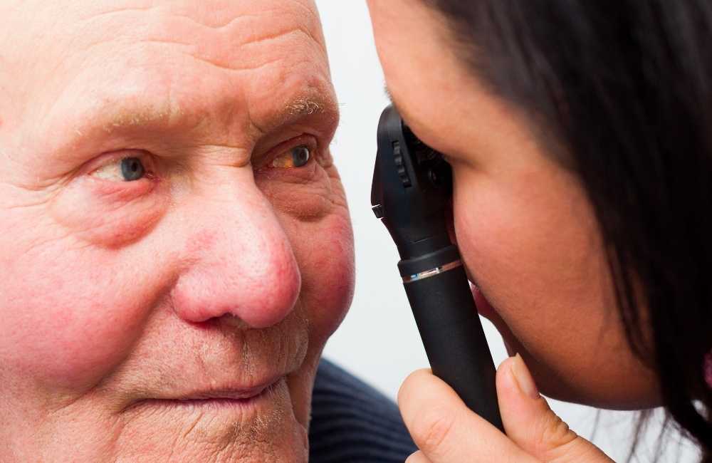
Diagnosis
-
Ophthalmoscopy
Diagnosis is best made with the pupil dilated. Well-developed cataracts appear as grey, white, or yellow-brown opacities in the lens. Examination of the red reflex through the dilated pupil with the ophthalmoscope held about 30 cm away usually discloses subtle opacities.
Small cataracts stand out as dark defects in the red reflex while large cataracts may obliterate the red reflex.
-
Slit-lamp examination
This provides more details about the character, location, and extent of the opacity.
Cataracts Treatment
-
Surgical removal of the cataract
Surgery is recommended when cataracts prevent one from going about his or her daily activities, such as reading, driving etc. It’s also performed when cataracts interfere with the treatment of other eye problems.
This is called extraction cataracts and is usually done using a topical or local anaesthetic and IV sedation. Two extraction techniques are deployed viz:
-
- Intracapsular cataract extraction
Here, the cataract and lens capsule is removed in one piece. This technique is rarely used.
-
- Extracapsular cataract extraction
-
-
- The hard central nucleus is removed in one piece and then the soft cortex is removed in multiple small pieces.
- Phacoemulsification (a type of extracapsular cataract extraction), involves the use of ultrasound waves to break the lens apart and remove the pieces.
-
Very small incisions are used, thus enabling the fastest healing, and is usually the preferred procedure.
-
- Femtosecond laser mode-locking
This is a technique by which a laser can be made to produce pulses of light of extremely short duration, on the order of picoseconds (10−12 s) or femtoseconds (10−15 s). A laser operating in this way is sometimes referred to as a femtosecond laser used, in modern cataract surgery.
-
Placement of an intraocular lens
A plastic or silicone lens is almost always implanted intraocularly (inside the eye) to replace the optical focusing power of the removed crystalline lens.
The lens implant is usually placed:
-
- On or within the lens capsule (posterior chamber lens).
- The lens can also be placed in front of the iris (anterior chamber lens).
- Or attached to the iris and within the pupil (iris plane lens).
Types of intraocular Lens
-
- Monofocal lenses
Monofocal lenses are set to maximize the patient’s best vision at one particular distance. Often, people choose to set them for distance vision and use reading glasses for near vision.
They are the most common type of lens used and are also covered by insurance in some countries.
-
- Toric lenses
These help to correct astigmatism.
-
- Extended depth–of–focus lenses
Extended depth-of-focus lenses have one corrective zone stretched to maximize the patient’s distance and intermediate vision. This is helpful for those using the computer and performing many types of work.
Post-surgical complications
Major complications of cataract surgery are rare. Complications include the following:
-
Intraoperative
-
- Bleeding beneath the retina, causing the intraocular contents to extrude through the incision (choroidal haemorrhage—very rare and could result in irreversible blindness.
- Vitreous prolapsing out of the incision (vitreous loss), fragments of the cataract dislocating into the vitreous.
- Incisional burns.
- Detachment of corneal endothelium and its basement membrane (Descemet membrane).
-
Within the first week
Endophthalmitis (infection within the eye) is very rare and could result in irreversible blindness and glaucoma
-
Within the first month
Cystoid macular oedema.
-
Months later
- Bullous keratopathy (i.e., swelling of the cornea due to damage to the corneal pump cells during cataract surgery).
- Retinal detachment and posterior capsular opacification (common but treatable with a laser)
Prevention of Cataracts
Many ophthalmologists recommend the following:
- Ultraviolet-coated eyeglasses or sunglasses as a preventive measure.
- Reducing risk factors such as alcohol, tobacco, and corticosteroids
- Controlling blood glucose in diabetes delay onset.
- A diet high in vitamin C, vitamin A, and carotenoids (contained in vegetables such as spinach and kale) may protect against cataracts.
Conclusion
Cataracts are the leading causes of blindness worldwide, which usually develop slowly over years and can be successfully managed surgically with a very good prognosis. Prioritize your eye health today, kindly see an eye care professional.