
Breast cysts are a common diagnosis among women and one of the most common reasons for a referral to a breast clinic. These cysts can be entirely asymptomatic and only discovered incidentally, or can be symptomatic, presenting as lumps, pain, or associated nipple discharge. Perhaps the most challenging are complicated cysts, that is, cysts with internal debris.
What are Breast Cysts?
Breast cysts are non-cancerous, fluid-filled sacs that are usually seen in the breast. Sometimes they can be felt, especially if they become painful a few days before the menstrual cycle. In most cases, breast cysts don’t need treatment.
In some women, breast cysts sometimes present with changes in fibrous connective tissue and glands in the breast. When presenting with pain and general lumpiness, fibrocystic changes are said to have taken place.
What Causes Breast Cysts?
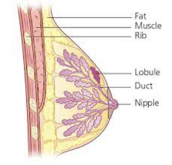 To understand breasts cysts, it’s important to have a little knowledge of the breast anatomy. Breasts are made up of lobules (milk-producing glands) and ducts (tubes that carry milk to the nipple).
The milk glands are surrounded by fibrous support tissue and fat, known as breast tissue. This tissue gives breasts their size and shape. Sometimes, the milk glands can fill up with fluid, these are breast cysts.
Cysts may appear naturally as the breast alters with age, due to normal changes in the estrogen hormone levels etc. During the menstrual cycle, estrogen causes fluid to be produced, although breast cysts can develop at any age, they’re most common in women over 35.
After the menopause (when your periods stop), as estrogen levels fall, cysts usually stop forming. Women who have hormone replacement therapy (HRT) may still get cysts.
Cysts begin when fluid starts to build up inside the breast glands. They start as microcysts (very small cysts), which are too small to feel unless they are part of a cluster (group) of microcysts. If fluid continues to build up, they can develop into macrocysts (large cysts). These can often be felt easily and can be as large as 1 or 2 inches across.
Types of Breast Cysts
Cysts can be simple, complex, or complicated:
-
Simple Breast Cysts
These cysts have smooth borders, thin walls, and are totally filled with fluid. They’re always benign.
-
Complex Breast Cysts
Unlike simple cysts, complex cysts have irregular borders, thick walls, and some solid matter within the fluid. Most are benign.
-
Complicated Breast Cysts
These cysts are somewhere in between simple and complex. They don’t have thick walls, but they may have some solid matter within the fluid. Most are benign.
What are the Symptoms of Breast Cysts?
Breast cysts can present in the following ways. Viz
- No symptoms at all.
- Breast Pain.
- Noticeable lump in the breast.
- Erythema- When a cyst is inflamed, erythema may be present at the site of the ‘‘lump,’’ and there may be associated tenderness.
Diagnosing Breast Cysts
Cysts usually become noticeable as a lump in the breast or are found by chance during a routine screening mammogram (breast x-ray) or while been investigated for another reason.
Diagnosis starts from viz
-
Physical Examination
After discussing your symptoms and health history, the doctor will physically examine the breast lump and check for any other breast abnormalities .A clinically detected breast mass cannot be characterised as a cyst on the basis of physical findings alone.
-
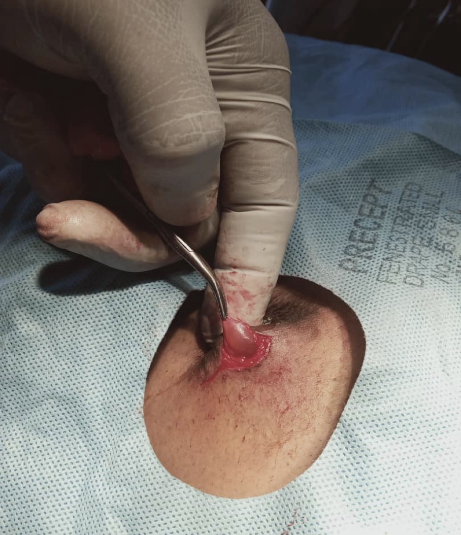
Needle Aspiration
If the lump can be easily felt, your specialist may put a fine needle into it and draw off the fluid to confirm that it’s a fluid-filled cyst. The fluid is examined under a microscope to check for cancer only if any of the following occurs:
- The fluid is bloody or cloudy.
- Little fluid is obtained.
- The lump is still present after the fluid is drained.
Otherwise, the woman is checked again in 4 to 8 weeks. If the cyst can no longer be felt at this time, it is considered noncancerous. If it has reappeared, it is drained again, and the fluid is examined under a microscope. If the cyst reappears a third time or if it is still present after it was drained, a biopsy is done.
-
Breast Ultrasound
If you’re under 40, you’re more likely to have an ultrasound than a mammogram. Younger women’s breast tissue is usually dense- this makes the x-ray image less clear, so normal changes or benign conditions can be harder to identify. However, for some women under 40 mammograms may still be needed to complete the assessment.When appropriate criteria are applied to a lesion,breast ultrasound is 96% to 100% accurate in the diagnosis of cysts.
-
Mammogram
This is an X-ray of the breast. usually, at least two pictures are taken of each breast, one breast at a time. The radiographer will position the breast firmly between two flat plates on the X-ray machine for up to a minute. This might feel uncomfortable.The mammographic appearance of cysts is variable. Cysts can occur as single or multiple, unilateral or bilateral masses of varying sizes and densities. Although many cysts are well circumscribed some have obscured or ill-defined margins. Spiculation or distortion is rare. Calcifications can develop in the cyst wall or within cysts (referred to as ‘‘milk of calcium’’). Unless associated with intra-cystic calcifications, most cysts are indistinguishable on mammography from other water density masses, including malignancies, and thus breast ultrasound is indicated for further evaluation.
-
Core Biopsy
If there is any question about the correct diagnosis, core biopsy is appropriate. If a core biopsy is undertaken, simple cysts decrease significantly in size and may disappear completely after the procedure. Histologically, pieces of epithelialcell– and apocrine cell–lined cysts are reported after core biopsy of simple cysts.
Other Fluid Collections
Other fluid collections seen in the breast include:
-
Postoperative or Traumatic Fluid Collections (e.g,hematomas)
Fluid collections in the breast may develop after surgery, particularly after removal of lump from the breasts (Lumpectomy) and radiation therapy or trauma. In some patients, fluid collections may result from trauma.
-
Galactoceles
Galactoceles are cysts that contain milky fluid and develop in women who are pregnant, lactating, or have stopped lactating within the last 2 to 3 years.
-
Abscesses
Patients who have breast abscesses can be divided into three groups:
- Patients who have infections related to breastfeeding,
- Patients who have recurring subareolar abscesses unrelated to nipple piercing or nipple rings.
- patients who have peripheral mastitis or abscesses unrelated to pregnancy orlactation.
The mammographic appearance of these lesions is variable. Round and oval masses with indistinct or ill-defined margins are common. Sonographically, these other fluid collections can appear as cystic, complex cystic, or, rarely, as solid masses.
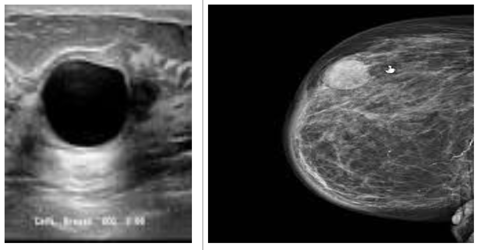
1. Breast cyst on Ultrasound 2. breast cyst on a Mammogram
Breast Cysts Treatment
No treatment is necessary for simple breast cysts — those that are fluid filled and don’t cause any symptoms — that are confirmed on breast ultrasound or after a fine-needle aspiration many cysts will disappear with no treatment. If a cyst persists, feels firmer or you notice skin changes on the skin over the cyst, follow up with your doctor.
-
Fine-Needle Aspiration (FNA)
Fine-needle aspiration may be used to diagnose and treat a breast cyst if all the fluid can be removed from the cyst during the procedure, and then your breast lump disappears and your symptoms resolve.
For some breast cysts, however, you may need to have fluid drained more than once. Recurrent or new cysts are common. If a breast cyst persists through two to three menstrual cycles and grows larger, see your doctor for further evaluation.
-
Hormone Use
Using birth control pills (oral contraceptives) to regulate your menstrual cycles may help reduce the recurrence of breast cysts. But because of possible significant side effects, birth control pills or other hormone therapy, such as tamoxifen, is usually recommended only for women with severe symptoms. Discontinuing hormone therapy after menopause may also help prevent breast cysts.
-
Surgery
Surgery to remove a breast cyst is necessary only in unusual circumstances. Surgery may be considered if an uncomfortable breast cyst recurs month after month or if a breast cyst contains blood-tinged fluid or shows other worrisome signs.
Conclusion
Breast cysts represent the most common cause of breast mass or breast symptoms in general. They are part of a larger benign disease process known as fibrocystic disease of the breast. This disease process is a wide spectrum of both fibrous and cystic changes in the breast tissue.
The simple breast cyst forms as an aberration in the natural breast development. The relationship between fibrocystic changes and breast cancer is complicated and controversial. Because of this, proper diagnosis, treatment, and management of breast cysts are essential.