Amebiasis occurs worldwide but is predominantly seen in developing countries with inadequate sanitation and increased fecal contamination of water supplies.
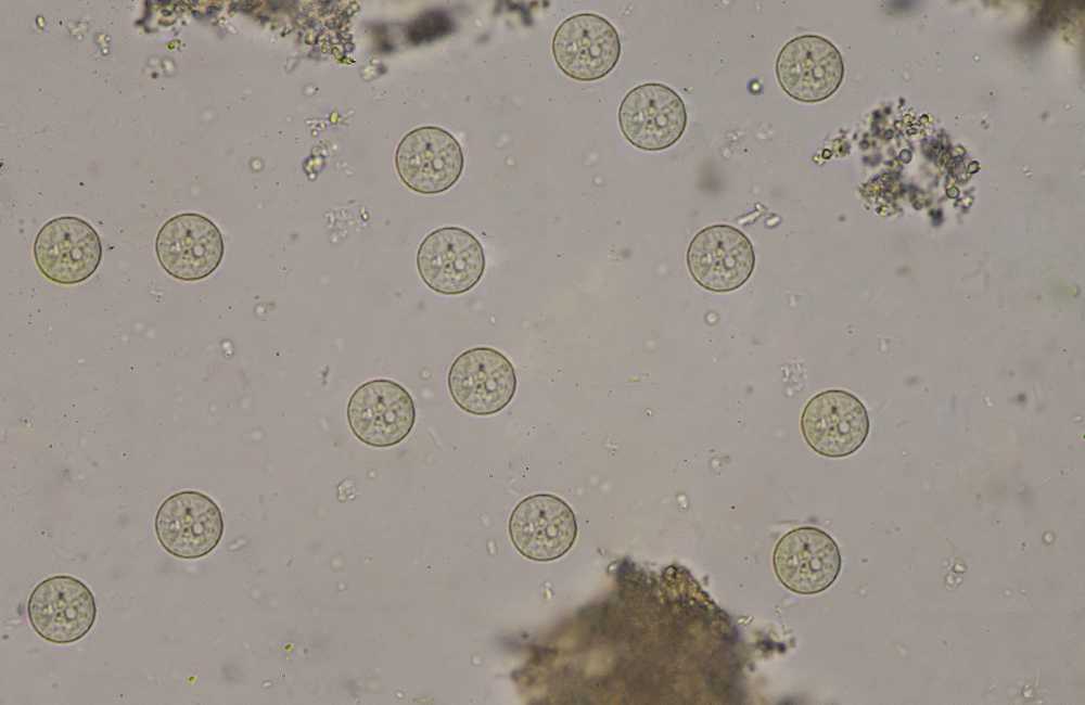
The principal source of infection is ingestion of water or food contaminated by feces containing Entamoeba histolytica cysts (E. Histolytica for short). Travelers to developing countries can acquire amebiasis when visiting endemic regions.
The organism E. histolytica is viable for prolonged periods in the cystic form in the environment. It can also be acquired after direct inoculation of the rectum, from anal or oral sex, or from equipment used for colonic irrigation. Despite the global public health burden, there are no vaccines or prophylactic medications to prevent amebiasis
What is Amebiasis?
Amebiasis is an infection of the large intestine and sometimes the liver or other organs caused by a single-cell protozoan parasite called Entamoeba histolytica. It is also called amebic dysentery.
Entamoeba species exist in two forms viz:
- An active parasite (trophozoite).
- A dormant parasite (cyst).
Transmission of Amebiasis
Infection begins when cysts are swallowed. The cysts hatch, releasing trophozoites that multiply and can cause ulcers in the lining of the intestine. Occasionally, they spread to the liver or other parts of the body.
Some trophozoites become cysts, which are excreted in stool (feces) along with trophozoites. Outside the body, the fragile trophozoites die. However, the hardy cysts can survive.
Cysts can be spread directly from person to person or indirectly through food or water. Amebiasis can also be spread through oral-anal sex.
Fruits and vegetables may be contaminated when grown in soil fertilized by human feces, washed in polluted water, or prepared by someone who is infected.
Amebiasis may occur and spread in places with adequate sanitation if infected people are incontinent or hygiene is poor (for example, in day care centers or psychiatric institutions).
Symptoms
Not everyone who has an amebiasis infection gets sick. One might not have any amebiasis symptoms, especially when first infected. Symptoms develop within four weeks after infection and they include:
- Cramping (abdominal pain).
- Diarrhea (sometimes with rectal bleeding).
- Fever.
- Watery loose stools.
- Nausea.
- Wasting of the body (emaciation) and anemia can occur in people with chronic infection.
- Sometimes large lumps (amebomas) may form inside the large intestine (colon) thus presenting as an abdominal mass.
- In some people, the amebas spread to the liver where they can cause an abscess (Liver abscess) with associated right upper abdominal pain or tenderness.
Investigations for Amebiasis
-
Stool tests (microscopy)
Diagnosis of E. histolytica traditionally, is by microscopic examination of the stool of the infected patient and this may require three to six stool samples to find the parasite, and even when they are seen, Entamoeba histolytica cannot be distinguished from some other related species like Entamoeba dispar, which looks hystolitica but is genetically different, does not cause disease.
-
Sometimes blood tests to detect antigens to the parasite or antibody reactions
-
- Antigen test
The best approach is to test the stool for a protein released by the Parasite (antigen testing) or to use the polymerase chain reaction (PCR) technique to check the stool for the parasites genetic material. The PCR technique produces many copies of its genetic material and thus makes the parasite easier to identify.
Antigen or PCR tests are more useful than microscopic examination of stool samples, which is often inconclusive.
The combination of serological tests with detection of the parasite (by antigen detection or PCR) offers the best approach to diagnosis.
-
- Antibody tests
Blood tests are then done to check for antibodies to the parasites. Antibodies are proteins produced by the immune system to help defend the body against a particular attack, including that by parasites.
Enzyme immunoassay (EIA) is the most widely used serologic test. Antibody titers can confirm E. histolytica infection although, it impossible to differentiate acute or current from past infection in residents from areas with a high prevalence of infection.
The EIA test detects antibody specific for E. histolytica in approximately 95% of patients with extra intestinal amebiasis, 70% of patients with active intestinal infection, and 10% of asymptomatic persons who are passing cysts of E. histolytica.
If antibodies are not detectable in patients with an acute presentation of suspected amebic liver abscess, a second specimen should be drawn 7-10 days later. If the second specimen does not show seroconversion, other agents should be considered. Detectable E. histolytica-specific antibodies may persist for years after successful treatment, so the presence of antibodies does not necessarily indicate acute or current infection.
Test for antibodies to E. histolytica should be done mostly by laboratories that can demonstrate technical expertise and understanding of the several serological tests that should be applied simultaneously with culture and PCR when extra-intestinal amebiasis is suspected.
-
- Colonoscopy
A flexible viewing tube (endoscope) may be used to look inside the large intestine. If ulcers or other signs of infection are found there, the endoscope is used to obtain a sample of fluid or tissue from the abnormal area.
-
- Imaging techniques
When amebas spread to sites outside the intestine (such as the liver), they may no longer be present in the stool. Ultrasonography, computed tomography (CT), or magnetic resonance imaging (MRI) can be done to confirm an abscess in the liver, but these tests do not indicate the cause.
Diagnosis
Diagnosis is from history, clinical suspicion and results of the investigations listed above. Amebic extraintestinal infection is more difficult to diagnose. Stool examination is usually negative, and recovery of trophozoites from aspirated pus is uncommon. If a liver abscess is suspected, ultrasonography, CT, or MRI should be done. They have similar sensitivity; however, no technique can differentiate amebic from pyogenic abscess with certainty.
Also in some situation especially if Liver abscess is suspected, drugs that kill Entamoeba (amebicide) is tried. If the person improves, the diagnosis is probably Amebiasis.
Prevention of Amebiasis
If traveling to places where the infection is common, the following regimen when preparing and eating food should be adhered to:
- Thoroughly wash fruits and vegetables before eating.
- Avoid eating fruits or vegetables unless you wash and peel them yourself.
- Use bottled water and soft drinks from sealed containers.
- If you must drink tap water boil it for at least 1 minute, or use a store-bought “absolute 1 micron” filter and add disinfecting chlorine, chlorine dioxide, or iodine tablets to the filtered water.
- Avoid ice cubes or fountain drinks.
- Avoid peeled fresh fruit or vegetables.
- Avoid milk, cheese, or other unpasteurized dairy products.
- Avoid food sold by street vendors.
Treatment for Amebiasis
-
Drugs
For gastrointestinal symptoms and extra-intestinal amebiasis, one of the following is used:
- Oral Metronidazole
-
- 500 to 750 mg 8hrly for 10 days (for adults).
- 12 to 17 mg/kg 8hrly for 10 days (in children.
- Tinidazole
-
- 2 g orally once/day for 3 days( for Adults)
- 50 mg/kg [maximum 2 g] orally once/day in children > 3 years) for 3 days for mild to moderate gastrointestinal symptoms, 5 days for severe gastrointestinal symptoms, and 3 to 5 days for amebic liver abscess.
These 2 drugs should not be taken by pregnant women. Alcohol must be avoided because these drugs have a disulfirian -like effect (Disulfiram works by providing a negative reaction if one drinks alcohol, used in the treatment of alcohol abuse. Symptoms of this reaction include nausea, vomiting, a racing heart, and flushing of the face or chest).
In terms of gastrointestinal adverse effects, tinidazole is generally better tolerated than metronidazole.
-
Fluid Therapy/supportive measures
Therapy for patients with significant gastrointestinal symptoms should include rehydration with fluid and electrolytes and other supportive measures.

The above listed drugs are not sufficient to eradicate cysts. Consequently, another oral drug is used to eradicate residual cysts in the intestine.
Options for cyst eradication are viz
- Iodoquinol
-
- 650 mg orally 8hrly for 20 days (taken after meals in adults).
- 10 to 13 mg/kg [maximum of 2 g/day] orally 8hrly for 20 days in children).
- Paromomycin
-
- 8 to 11 mg/kg orally 8hrly for 7days (taken after meals).
Complications
Complications of Amebiasis may include:
- Liver abscess (collection of pus in the liver).
- Medicine side effects, including nausea and vomiting.
- Spread of the parasite through the blood to the liver, lungs, brain, or other organs.
- Dissemination in the brain.
- Bowel perforation.
- Stricture of the colon.
- Gastrointestinal bleeding.
- Empyema.( collection of pus in the space between the lung and the inner surface of the chest wall (pleural space).
Conclusion
Amebiasis is a relatively common parasitic infection. An important component of treatment is patient education. All travelers should maintain good personal hygiene, sanitation, and avoid high-risk sexual practices.
The E. histolytica cysts are relatively resistant to disinfection of water with chlorine. Drinking boiled or bottled water is advised.
All food should be washed, and the skin of fruits should be peeled. If abdominal pain symptoms, cramps, and diarrhea persist, a visit to the healthcare provider is advised.