
Acute pancreatitis is said to result when the pancreas gets severely inflamed. The pancreas is about 6 inches long, prismoid-shaped accessory digestive organ that lies transversely in the upper abdomen between the duodenum on the right and the spleen on the left.
Accessory organs are organs which are not part of the digestive system but aid in the digestion process by performing many secondary functions.
It is divided into head, neck, body and tail with the organ being entirely retroperitoneal with the exception of the tail that is intraperitoneal. The head of the pancreas, the enlarged part of the gland, is surrounded by the C-shaped curve of the duodenum.
Here, the main pancreatic duct carrying the pancreatic secretions joins with bile duct to form hepatopancreatic ampulla, which opens into the descending part of the duodenum.
The hepatopancreatic sphincter of Oddi around the hepatopancreatic ampulla is a smooth muscle sphincter that controls the flow of bile and pancreatic juice into the ampulla and inhibits reflux of duodenal substances into the ampulla.
The pancreas is a gland that does 2 main things viz:
- Makes enzymes that help break down food and send them into your small intestine.
- Also makes the hormones (insulin and glucagon) that control the body’s blood sugar levels.
What is Acute Pancreatitis?
Acute pancreatitis is usually a sudden and severe inflammation of the pancreas sometimes involving adjacent tissues, with gallstones and alcohol intake as the most common triggers (accounting for about 70 % of cases).
Acute pancreatitis is classed as mild, moderately sever and sever based on the presence of local complications and transient or persistent organ failure.
There are 2 types of acute pancreatitis viz
- Interstitial pancreatitis
Most patients develop this type of pancreatitis and it is self-limiting and is defined by the presence of an enlarged pancreas on imaging. Peripancreatic stranding may be seen and this signifies inflammation.
- Necrotizing pancreatitis
This is defined by the presence of pancreatic and/or peripancreatic necrosis. It is best seen on contrast-enhanced cross-sectional imaging.
Causes
- Gallstones.
This is the most common cause of acute pancreatitis. It is believed that increased pressure in the pancreatic duct (ductal hypertension) following stone obstruction at the ampulla or edema caused by passage of a stone may be responsible for aberrant activation of digestive enzymes and consequent activation of inflammatory cascade pathway.
- Alcohol intake.
This is another common cause of acute pancreatitis. The risk of developing pancreatitis increases with increased dose of the alcohol ingested.
The pancreatic acinar cells metabolize alcohol into a toxic metabolite, thus predisposing the cells to autodigestive injury, pancreatic necrosis, inflammation and cell death.
- Autosomal dormant mutation of cationic trypinogen gene
This is known to cause acute pancreatitis in about 80% of carriers and is also implicated in cystic fibrosis.
Signs of Acute pancreatitis
- Abdominal pain.
Pain is often the dominant symptom in both acute and chronic pancreatitis. Acute pancreatitis attack causes steady, boring upper abdominal pain that radiates through to the back in about 50% of patients.
This is usually of sudden onset especially in gallstone pancreatitis. In alcoholic pancreatitis, the pain develops over a few days and usually persists for several days. Coughing, vigorous movement, and deep breathing may accentuate it.
-
- Nausea.
- Vomiting.
- Rapid heart rate.
The patient tends to appear acutely ill and sweaty. Pulse rate is usually elevated.
- Breathing difficulties
The lungs may have limited diaphragmatic excursion and evidence of atelectasis (complete or partial collapse of the lungs)
- Fever.
Temperature may be normal or even subnormal at first but may increase to 37.7 -38.3°C within a few hours.
- Swelling and feeling sore or tender in your upper belly
- Fluid buildup in the abdomen.
Pancreatic duct disruption may cause ascites (pancreatic ascites). Infection in the pancreas or in an adjacent fluid collection should be suspected if the patient has a generally toxic appearance with fever and an elevated white blood cell count or if deterioration follows an initial period of stabilization.
- Lowered blood pressure
Blood pressure may be transiently high or low, with significant postural hypotension.
- Yellowing of the skin and eyes (jaundice).
Scleral icterus is occasionally present because of obstruction of the bile duct by a gallstone or inflammation and swelling of the pancreatic head.
- Blunted sensorium.
Sensorium may be blunted to the point of obtundation (reduced level of alertness or consciousness).
- Paralytic ileus
Patients may have an ileus resulting in decreased bowel sounds and abdominal distention.
- Extravasation of hemorrhagic exudate.
The grey turner sign (discoloration of the skin resulting from bleeding underneath at the flanks) and the Cullen sign (discoloration of the skin resulting from bleeding underneath at the umbilical region) indicate extravasation of hemorrhagic exudate, occur in less than 1% of cases, and portend a poor prognosis.
- Multiorgan failure.
Patients with severe disease can develop multiorgan failure (cardiovascular, renal, and respiratory).
Investigating Acute Pancreatitis
-
Laboratory investigations
-
- Serum markers (amylase, lipase)
Serum amylase and lipase concentrations increase on the first day of acute pancreatitis and return to normal in 3 to 7 days.
Amylase and lipase are 2 enzymes produced by the pancreas. Elevations in lipase are generally considered a better indicator for pancreatitis as it has greater specificity and has a longer half-life. Both enzymes may also be increased other conditions viz:
- In renal failure.
- Various abdominal conditions (e.g. perforated ulcer, mesenteric vascular occlusion, intestinal obstruction) etc.
- Salivary gland dysfunction.
- Macroamylasemia (a condition when amylase is combined with a protein molecule thus creating a very large molecule making kidney excretion very difficult and slow).
- Tumors that secrete amylase.
Fractionation or separation of total serum amylase into pancreatic types (p-type) isoamylase and salivary-type (s-type) isoamylase increases the accuracy of serum amylase.
Please note that both amylase and lipase levels may remain normal if acinar tissue were previously destroyed during previous episodes. This precludes release of sufficient amounts of enzymes.
-
- Complete blood count
The white blood cell count usually increases to 12,000 to 20,000/mcL (12 to 20 × 109/L) and the hematocrit to as high as 50 to 55% due to third-space fluid losses.
-
- Electrolytes/urea/creatinine
Raised blood urea nitrogen is indicative of severe inflammation. Persistent elevation in BUN despite resuscitation is an indicator of increased morbidity and mortality.
Patients with shock may have an elevated anion gap metabolic acidosis or other electrolyte abnormalities.
-
- Blood sugar levels
High blood sugar levels (hyperglycemia) and low calcium levels(hypocalcemia) may occur.
-
- Liver function tests
Patients may have abnormal liver function test results, including elevated serum bilirubin, due to a retained stone in the bile duct or compression of the bile duct by pancreatic edema.
-
- Urine dipstick test for trypsinogen-2
A urine dipstick test for trypsinogen-2 has sensitivity and specificity of > 90% for acute pancreatitis
-
Imaging studies
-
- Chest x-ray
Should be done and may reveal lung collapse or a fluid in the lungs (pleural effusion). These are signs of severe disease.
-
- Plain abdominal x-ray
Plain x-rays of the abdomen may disclose calcifications within pancreatic ducts (evidence of prior inflammation. Hence chronic pancreatitis), calcified gallstones, localized ileus of a segment of small intestine in the left upper quadrant or the center of the abdomen or there may be absence of air in left colonic flexure or descending colon in more severe disease.
However, the value of routine abdominal x-rays is controversial.
-
- Ultrasound scan
Should be done if gallstone pancreatitis is suspected (and another cause is not obvious) to detect gallstones or dilation of the common bile duct, which indicates biliary tract obstruction. Edema of the pancreas may be visible, but overlying gas frequently obscures the pancreas.
-
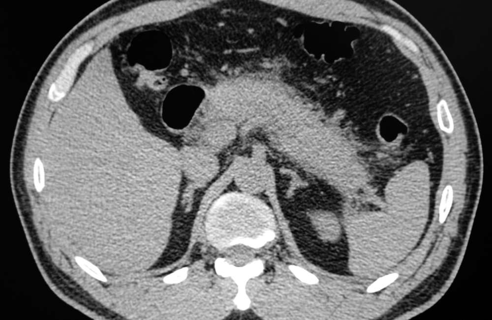
- CT-with intravenous contrast
CT with an intravenous contrast is the imaging study of choice to establish the diagnosis of acute pancreatitis and to assess for local complications.
Diagnosis of Acute Pancreatitis
Acute pancreatitis is suspected whenever severe unexplained abdominal pain occurs, especially in a patient with significant alcohol use or known gallstones.
The diagnosis of acute pancreatitis is established by the presence of at least 2 of the following:
- Abdominal pain consistent with the disease.
- Serum amylase and/or lipase that is 3 times more than the upper limit of normal (normal range of amylase and lipase levels can differ depending on the assay used).
- Characteristic findings on contrast-enhanced cross-sectional imaging studies.
Severity Of Acute Pancreatitis
Severity of acute pancreatitis can be classified as
- Mild
In mild acute pancreatitis, inflammation is confined to the pancreas and its close vicinity. Patients do not have organ failure or systemic or local complications. Mortality is rare.
- Moderately severe
In moderately severe acute pancreatitis, patients have local or systemic complications but no organ failure, or only transient organ failure (resolves within 48 hours).
- Severe
In severe acute pancreatitis, there is persistent single or multiorgan failure (more than 48 hours). Most patients have one or more local complications. The mortality rate is more than 30%.
Treatment Of Acute Pancreatitis
Treatment of acute pancreatitis is typically supportive. Patients who develop complications may require specific additional treatment.
-
Supportive measures
-
- Early goal-directed fluid resuscitation.
Early, aggressive fluid resuscitation, defined as 250 to 500 mL/hour of isotonic crystalloid solution (ideally lactated Ringer’s solution), be provided to all patients during the first 12 to 24 hours unless contraindicated by cardiovascular, renal, or other related comorbid factors.
Adequacy of fluid replacement can be assessed by reduction in hematocrit and blood urea nitrogen levels over the first 24 hours, particularly if they were high at the onset.
-
- Analgesia
Adequate pain relief requires use of parenteral opioids such as hydromorphone or fentanyl, which should be given in adequate doses. Morphine can theoretically increase pressure in the sphincter of Oddi, thus making hydromorphone preferable to morphine.
-
- Antiemetic (for vomiting)
Antiemetic drugs should also be given to relieve nausea and vomiting.
-
- Oxygen
Because pancreatitis can cause lung injury and affect normal lung function, supplemental oxygen is occasionally delivered through breathing tubes that are connected via the nose (e.g., nasal cannula) or via a mask.
-
- Nutritional support
Early enteral nutrition is recommended because it is associated with lower morbidity compared to delay or no nutrition. Patients with mild pancreatitis can begin an oral low-residue, low fat, soft diet as soon as it can be tolerated.
Enteral feeding is preferred over total parenteral nutrition because parenteral nutrition is associated with increased risk of infectious complications and organ failure.

-
Antibiotic treatment
For severe acute pancreatitis with complications, antibiotics and therapeutic interventions are needed. The management of patients with severe acute pancreatitis and its complications should be individualized using a multidisciplinary approach including therapeutic endoscopists, interventional radiologists, and a surgeon and should be monitored closely in the first 24 to 48 hours n an ICU.
Complications Of Acute Pancreatitis
Complications of acute pancreatitis can be categorized viz
-
Local
-
- Pancreatic and peripancreatic fluid collections.
Enzyme-rich pancreatic fluid collection can occur early in the disease course in the pancreas or around it. These collections may contain only fluid or in some cases necrotic materials and they seem to resolve spontaneously, but if they do not resolve after about 4 weeks and go on to develop fibrous capsule, the collection is now termed pancreatic pseudocysts and they can also resolve spontaneously or becomes infected by gut bacteria thus leading to very high morbidity and mortality.
-
- Splenic vein thrombosis.
Occurs when blood clots block veins in the spleen.
-
- Pseudo aneurysm formation.
A pseudoaneurysm occurs when a blood vessel wall is injured. Blood leaking from the vessel collects in surrounding tissue. It is sometimes called a false aneurysm.
-
- Gastric outlet dysfunction.
Patients with acute pancreatitis can experience gastric outlet dysfunction or obstruction as a result of narrowing of the duodenum or pylorus caused by surrounding pancreatic and peripancreatic inflammation. It can also be the result of compression by walled-off necrosis or pseudocyst.
-
Systemic
-
- Shock
In severe cases, parts of the pancreas die, a condition referred to as necrotizing pancreatitis. This can cause pancreatic fluid and blood to leak into the abdominal cavity, decreasing the blood volume and blood pressure thus leading to hypovolemic shock.
-
- Organ failure
Here, organ failure can be a single organ or multiple organs shut down (e.g., cardiovascular and/or respiratory failure, acute kidney injury).
Organ failure is defined using a scoring system, which is based on laboratory and vital sign indicators of respiratory, renal, and cardiovascular impairment.
Risk is increased in patients with underlying co-morbidities and/or a persistent systemic inflammatory response syndrome (SIRS).
Systemic inflammatory response syndrome (SIRS), in the appropriate clinical setting, is defined as the presence of two or more of the following:
- Temperature of more than 38.3° C or less than 36.0° C
- Heart rate of more than 90/minute
- Respiratory rate more than 20cycles/minute or PACO2 less than 32 mmHg.
- White blood cell counts of more than 12,000/mcL (12 × 109/L), less than 4,000/mcL (4 × 10⁹/L) or with more than 10% bands
Conclusion
Medical treatment of mild acute pancreatitis is relatively straightforward. It is important that patients with severe acute pancreatitis are identified as soon as possible because this is critical for achieving optimal outcomes.
Management depends largely on severity. Treatment of severe acute pancreatitis involves intensive care while surgical intervention (open or minimally invasive) is indicated in selected cases.